Female Urogenital System XC-332
₹2,427.00 Inclusive all Taxes
-
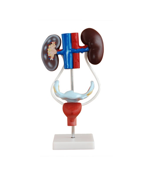
This model shows kidney, ureters, urinary bladder, uterus, accessories of uterus, vagina, ovary membrane, ligaments, uterus ligament and its artery etc. Made of PVC plastic.
- Made of Washable Wand Unbreakable PVC Plastic
- Superior quality that closely resembles to orignal skeleton
In stock
Female Urogenital System is a specialized and highly detailed model designed to replicate the female urinary and reproductive anatomy. It serves as a valuable tool in medical education and clinical practice, offering an anatomically accurate representation of the internal structures involved in both urinary and reproductive functions.
Anatomy of the Female Urogenital System XC-332:
The XC-332 model includes the essential components of the female urogenital system, incorporating both the urinary tract and the reproductive system. It highlights the key structures and their interrelations, providing a comprehensive understanding of female anatomy.
- Urinary System:
- Kidneys: The model features a pair of kidneys that filter waste from the bloodstream and produce urine. These organs are responsible for maintaining fluid and electrolyte balance in the body.
- Ureters: The model accurately represents the two ureters, which transport urine from the kidneys to the bladder. These tubular structures have smooth muscle layers that contract rhythmically to propel urine.
- Bladder: The bladder is depicted as a hollow, muscular organ that stores urine until it is expelled from the body. The model provides a clear view of its internal lining and how it adapts to changes in volume.
- Urethra: The urethra, which connects the bladder to the outside world, is also included. This structure serves as the conduit for urine to exit the body.
- Reproductive System:
- Ovaries: The XC-332 model showcases the ovaries, which are responsible for producing eggs (ova) and hormones such as estrogen and progesterone. It provides a clear view of their location and structural features.
- Fallopian Tubes: The fallopian tubes are depicted as the pathways through which eggs travel from the ovaries to the uterus. The model highlights their important role in fertilization, where sperm meets the egg.
- Uterus: The model features the uterus, a pear-shaped organ where fetal development occurs. The uterine walls are represented in detail, showing the endometrium (inner lining), myometrium (muscular layer), and serosa (outer layer). The model provides a realistic view of the uterus’ ability to expand during pregnancy.
- Cervix: The cervix, the lower part of the uterus, is depicted as the gateway between the uterus and the vaginal canal. The model illustrates its structure, including the cervical canal, which is important for the passage of menstrual fluid, sperm, and the birth of a baby.
- Vagina: The vagina is shown as a muscular canal that connects the cervix to the outside of the body. The model provides a realistic depiction of the vaginal walls and their role in sexual intercourse and childbirth.
Functionality and Utility:
The XC-332 model is designed with a high level of anatomical precision, making it suitable for a variety of educational and clinical applications. It offers an opportunity for students, healthcare professionals, and patients to gain a clear understanding of the anatomy and functions of the female urogenital system.
Medical students can use the model to study the structures of the female urinary and reproductive systems in a detailed and hands-on manner. It aids in learning the relationships between organs, the physiological processes involved in urination and reproduction, and common pathologies affecting these systems.
For healthcare providers, such as gynecologists and urologists, the XC-332 model serves as an excellent tool for explaining complex concepts to patients, especially when discussing conditions like urinary incontinence, reproductive health issues, or pelvic floor disorders.
Material and Design:
Constructed from durable, high-quality materials, the XC-332 model is both realistic and long-lasting. The model’s surface mimics human tissue, offering a lifelike appearance and tactile experience for learners. The clear labeling and attention to detail make it easy to identify each structure, enhancing its educational value.
In conclusion, the Female Urogenital System XC-332 is an advanced anatomical model that serves as a vital resource for medical education, patient communication, and clinical practice. Its intricate design allows users to study the female urinary and reproductive systems in an interactive and comprehensive manner.
| Weight | 14 kg |
|---|---|
| Dimensions | 75 × 38 × 40 cm |
| Age | |
| Assembly Required | No |
| Batteries Included | No |
| Batteries Required | No |
| Color | MultiColor |
| Country of Origin | Made In India |
| Gender | Unisex |
| HSN Code | 9503 |
| Remote Controlled Included | No |
Only logged in customers who have purchased this product may leave a review.
Related products
Scientific Models
Scientific Models
Scientific Models
Scientific Models








Reviews
There are no reviews yet.