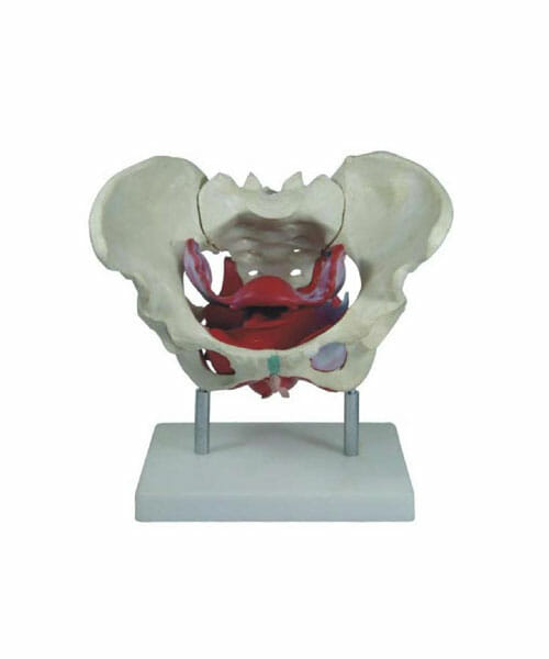
Female Pelvis Muscles and Organs XC-125
₹3,398.00 Inclusive all Taxes
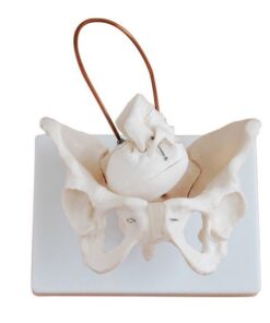
The Female Pelvis Muscles and Organs XC-125 model typically refers to an anatomical representation used for educational and medical purposes. Here’s a description of such a model:
Description:
The Female Pelvis Muscles and Organs XC-125 is a high-detail anatomical model designed to showcase the structure and spatial relationships of the pelvic organs and surrounding musculature. It is ideal for medical education, patient demonstrations, and anatomy studies.
Features:
- Pelvic Organs:
- Includes the uterus, ovaries, fallopian tubes, vagina, bladder, and rectum.
- Demonstrates the positioning and relationships between these organs.
- Often features removable parts for detailed examination.
- Pelvic Floor Muscles:
- Illustrates the levator ani group (pubococcygeus, iliococcygeus) and coccygeus muscles.
- Displays the role of the muscles in supporting pelvic organs and controlling bodily functions.
- Bone Structures:
- Incorporates the pelvic girdle (ilium, ischium, and pubis) for context and anatomical reference.
- May include the sacrum and coccyx to show how they anchor pelvic muscles.
- Vascular and Nervous System Details:
- Some models include representations of pelvic blood vessels and nerves, such as the internal iliac arteries and pudendal nerve, for advanced learning.
- Material:
- Typically constructed from durable, medical-grade materials.
- Painted or labeled to differentiate muscles, organs, and structures.
Applications:
- Medical Training: Aids in teaching gynecological, urological, and obstetric procedures.
- Patient Education: Helps explain conditions like pelvic floor dysfunction, incontinence, or prolapse.
- Research and Study: Supports in-depth anatomical studies in medical schools and clinics.
Would you like more information about the model’s specific components or potential usage scenarios?
Only 2 left in stock






Reviews
There are no reviews yet.