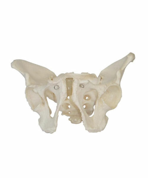
Adult Male Pelvis XC-123
₹2,427.00 Inclusive all Taxes
- The size of this model is just the same as the realities and made of PVC plastic.
It shows the following:
Pelvis shapes: Long and narrow
Pelvis cava: Like a funnel
Aperture pelvis superior: Heart-like
Sacrum: Long and narrow with great flexibility
Angles of pelvis arch:70-75 degree
Symphysis pubica: Long and narrow
Only 2 left in stock
Adult Male Pelvis model shows the structure of a male pelvis in life-size dimensions. The special plastic used to construct the model ensures realistic surface structure and colour. The structures illustrated include the pubis with pubic arch and pubic symphysis, obturator foramen, ischium, acetabulum, spina iliaca anterior, ilium, iliac crest, sacroiliac joint and the sacrum.
This model pelvis represents the size of an average adult male. A great value model for demonstration of the basic anatomy of the male pelvis.
The male pelvis shows the following typical features, in contrast to the female pelvis:
The Adult Male Pelvis XC-123 is a highly detailed anatomical model designed to provide a realistic and accurate representation of the adult male pelvis for educational, clinical, and research purposes. It serves as an essential tool for professionals in medical fields such as anatomy, surgery, orthopedics, radiology, and physiotherapy, as well as students and educators in healthcare programs. The model is crafted to depict the bones, joints, and key anatomical features of the adult male pelvis, offering a thorough understanding of its structure and function.
The XC-123 Pelvis is typically made from high-quality, durable materials, such as medical-grade plastic or resin, ensuring long-lasting use and the ability to withstand repeated handling. The model is often mounted on a sturdy base, allowing for easy display and interaction. It features lifelike detailing of the pelvic bones, including the iliac crests, sacrum, coccyx, pubic symphysis, and the acetabulum. These structures are presented with precise dimensions, ensuring accuracy and fidelity to human anatomy.
One of the key aspects of the Adult Male Pelvis XC-123 is its representation of the pelvic joints. The model clearly illustrates the sacroiliac joints, the pubic symphysis, and the hip joints, making it an invaluable tool for understanding the biomechanics and movement of the pelvis. This feature is particularly helpful for students and practitioners learning about gait, posture, and the role of the pelvis in weight-bearing activities.
In addition to its detailed skeletal features, the XC-123 model also includes visual markers or removable parts to enhance the learning experience. Some models may have detachable parts such as the femur, the pelvic inlet, or the sacrum to allow for further study and exploration of the pelvic region’s anatomy. Some advanced versions of the model include a cross-sectional view of the pelvis, showing muscles, arteries, veins, and nerves within the pelvic cavity, providing a more comprehensive learning experience.
For professionals in the field of radiology and imaging, the Adult Male Pelvis XC-123 may be used to simulate various diagnostic procedures, such as X-rays or CT scans, enabling practitioners to understand how different imaging modalities capture the structures of the pelvis. The model’s clarity and detail make it an excellent teaching aid in demonstrating fractures, degenerative changes, and other pathologies of the pelvic region.
The Adult Male Pelvis XC-123 is also beneficial for medical students and educators as it supports practical learning methods, helping individuals to visualize and palpate anatomical structures in a way that goes beyond theoretical studies. The model can be used for simulations of surgical procedures or for preparing patients for pelvic surgery, offering them a realistic understanding of the area being treated.
Overall, the Adult Male Pelvis XC-123 provides a highly accurate, durable, and versatile educational tool that facilitates a deep understanding of pelvic anatomy. It is ideal for use in classrooms, clinics, hospitals, and medical laboratories, assisting students, educators, and healthcare professionals in mastering the complex structure and function of the male pelvis.
| Weight | 14 kg |
|---|---|
| Dimensions | 82 × 44 × 33 cm |
| Age | |
| Assembly Required | No |
| Batteries Included | No |
| Batteries Required | No |
| Color | White |
| Country of Origin | Made In India |
| Gender | Unisex |
| HSN Code | 9503 |
| Remote Controlled Included | No |
Only logged in customers who have purchased this product may leave a review.
Related products
Scientific Models
Medium Skeleton with Nerves and Blood Vessels 85cm tall XC-102B
Scientific Models
Scientific Models
Scientific Models
Scientific Models
Scientific Models





Reviews
There are no reviews yet.